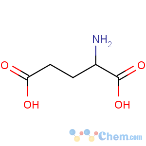
D-Glutamic Acid
-
- Product NameD-Glutamic Acid
- CAS No.138-16-9
- Purity99%
- Min Quantity
- Price0~100

 View Contact Detail
-
 Molecular Structure

- D-Glutamic Acid
Detailed Description
D-Glutamic Acidproduct Name: D-Glutamic Acid
Synonyms: D-2-Aminoglutaric acid; D-Glutaminic acid; D(-)-Glutamic acid; D-(-)-Glutamic acid; (2R)-2-ammoniopentanedioate
Molecular Formula: C5H8NO4
Molecular Weight: 146.1219
CAS Registry Number: 6893-26-1;138-16-9
EINECS: 230-000-8
Packing:25kg/drum
Assay:98%
Glutamic acid Information
------------------------------------------
Glutamic acid (abbreviated as Glu or E) is one of the 20-23 proteinogenic amino acids, and its codons are GAA and GAG. It is a non-essential amino acid. The carboxylate anions and salts of glutamic acid are known as glutamates. In neuroscience, glutamate is an important excitatory neurotransmitter that plays the principal role in neural activation.
Chemistry
-------------------------------------------
The side chain carboxylic acid functional group has a pKa of 4.1 and therefore exists almost entirely in its negatively charged deprotonated carboxylate form at pH values greater than 4.1; therefore, it is negatively charged at physiological pH ranging from 7.35 to 7.45.
Function and uses
---------------------------------------------
Metabolism
Glutamate is a key compound in cellular metabolism. In humans, dietary proteins are broken down by digestion into amino acids, which serve as metabolic fuel for other functional roles in the body. A key process in amino acid degradation is transamination, in which the amino group of an amino acid is transferred to an α-ketoacid, typically catalysed by a transaminase.
Ammonia (as ammonium) is then excreted predominantly as urea, synthesised in the liver. Transamination can thus be linked to deamination, effectively allowing nitrogen from the amine groups of amino acids to be removed, via glutamate as an intermediate, and finally excreted from the body in the form of urea.
Glutamate is also a neurotransmitter , which makes it one of the most abundant molecules in the brain. Malignant brain tumors known as glioma or glioblastoma exploit this phenomenon by using glutamate as an energy source, especially when these mutations become more dependent on glutamate due to mutations in the gene IDH1.
Neurotransmitter
Glutamate is the most abundant excitatory neurotransmitter in the vertebrate nervous system. At chemical synapses, glutamate is stored in vesicles. Nerve impulses trigger release of glutamate from the presynaptic cell. Glutamate acts on ionotropic and metabotropic (G-protein coupled) receptors. In the opposing postsynaptic cell, glutamate receptors, such as the NMDA receptor or the AMPA receptor, bind glutamate and are activated. Because of its role in synaptic plasticity, glutamate is involved in cognitive functions such as learning and memory in the brain. The form of plasticity known as long-term potentiation takes place at glutamatergic synapses in the hippocampus, neocortex, and other parts of the brain. Glutamate works not only as a point-to-point transmitter, but also through spill-over synaptic crosstalk between synapses in which summation of glutamate released from a neighboring synapse creates extrasynaptic signaling/volume transmission. In addition, glutamate plays important roles in the regulation of growth cones and synaptogenesis during brain development as originally described by Mark Mattson.
Glutamate transporters, EAAT and VGLUT, are found in neuronal and glial membranes. They rapidly remove glutamate from the extracellular space. In brain injury or disease, they can work in reverse, and excess glutamate can accumulate outside cells. This process causes calcium ions to enter cells via NMDA receptor channels, leading to neuronal damage and eventual cell death, and is called excitotoxicity. The mechanisms of cell death include
★Damage to mitochondria from excessively high intracellular Ca2+
★Glu/Ca2+-mediated promotion of transcription factors for pro-apoptotic genes, or downregulation of transcription factors for anti-apoptotic genes
Excitotoxicity due to excessive glutamate release and impaired uptake occurs as part of the ischemic cascade and is associated with stroke,autism[citation needed], some forms of intellectual disability, and diseases such as amyotrophic lateral sclerosis, lathyrism, and Alzheimer's disease. In contrast, decreased glutamate release is observed under conditions of classical phenylketonuria leading to developmental disruption of glutamate receptor expression.
Glutamic acid has been implicated in epileptic seizures. Microinjection of glutamic acid into neurons produces spontaneous depolarisations around one second apart, and this firing pattern is similar to what is known as paroxysmal depolarizing shift in epileptic attacks. This change in the resting membrane potential at seizure foci could cause spontaneous opening of voltage-activated calcium channels, leading to glutamic acid release and further depolarization.
Experimental techniques to detect glutamate in intact cells include using a genetically engineered nanosensor.The sensor is a fusion of a glutamate-binding protein and two fluorescent proteins. When glutamate binds, the fluorescence of the sensor under ultraviolet light changes by resonance between the two fluorophores. Introduction of the nanosensor into cells enables optical detection of the glutamate concentration. Synthetic analogs of glutamic acid that can be activated by ultraviolet light and two-photon excitation microscopy have also been described.This method of rapidly uncaging by photostimulation is useful for mapping the connections between neurons, and understanding synapse function.
Evolution of glutamate receptors is entirely the opposite in invertebrates, in particular, arthropods and nematodes, where glutamate stimulates glutamate-gated chloride channels.The beta subunits of the receptor respond with very high affinity to glutamate and glycine.Targeting these receptors has been the therapeutic goal of anthelmintic therapy using avermectins.
Brain nonsynaptic glutamatergic signaling circuits
Extracellular glutamate in Drosophila brains has been found to regulate postsynaptic glutamate receptor clustering, via a process involving receptor desensitization. A gene expressed in glial cells actively transports glutamate into the extracellular space,while, in the nucleus accumbens-stimulating group II metabotropic glutamate receptors, this gene was found to reduce extracellular glutamate levels.This raises the possibility that this extracellular glutamate plays an "endocrine-like" role as part of a larger homeostatic system.
GABA precursor
Glutamate also serves as the precursor for the synthesis of the inhibitory gamma-aminobutyric acid (GABA) in GABA-ergic neurons. This reaction is catalyzed by glutamate decarboxylase (GAD), which is most abundant in the cerebellum and pancreas.
Flavor enhancer
Main article: Glutamic acid (flavor)
Glutamic acid, being a constituent of protein, is present in every food that contains protein, but it can only be tasted when it is present in an unbound form. Significant amounts of free glutamic acid are present in a wide variety of foods, including cheese and soy sauce, and is responsible for umami, one of the five basic tastes of the human sense of taste. Glutamic acid is often used as a food additive and flavor enhancer in the form of its salt, known as monosodium glutamate (MSG).
Nutrient
All meats, poultry, fish, eggs, dairy products, and kombu are excellent sources of glutamic acid. Some protein-rich plant foods also serve as sources. 30% to 35% of the protein in wheat is glutamic acid. Ninety-five percent of the dietary glutamate is metabolized by intestinal cells in a first pass
Plant growth
Auxigro is a plant growth preparation that contains 30% glutamic acid.
NMR spectroscopy
In recent years, there has been much research into the use of